מיליה
נבדק על ידי ד"ר היילי וילאסי, FRCGP עודכן לאחרונה על ידי Dr Colin Tidy, MRCGPעודכן לאחרונה 14 דצמבר 2022
עומד בהנחיות העריכה של Patient
- הורדהורד
- שתף
- Language
- דיון
- Ask Patient
- גרסת שמע
- הוסף למקורות מועדפים בגוגל
אנשי מקצוע רפואיים
מאמרי עיון מקצועיים מיועדים לשימוש של אנשי מקצוע בתחום הבריאות. הם נכתבים על ידי רופאים מבריטניה ומבוססים על ראיות מחקריות, הנחיות בריטיות ואירופאיות. ייתכן שתמצא את מיליה מאמר מועיל יותר, או אחד מהאחרים שלנו מאמרי הבריאות.
What are milia?
Milia are very common, benign, keratin-filled epidermoid cysts that occur in individuals of all ages, from infants to elderly persons.1 .
Primary milia arise spontaneously and are typically seen in infants, when they are congenital, but may also occur in children and adults. Primary milia arise on skin bearing vellus hair follicles, most commonly on the face.
Secondary milia result from damage to eccrine ducts.
Milia en plaque and multiple eruptive milia are distinct entities.
How common are milia?1
Milia are common in all ages and both sexes. Congenital milia affect approximately 50% of infants.
Multiple eruptive milia and milia en plaque are extremely rare.
Milia symptoms2
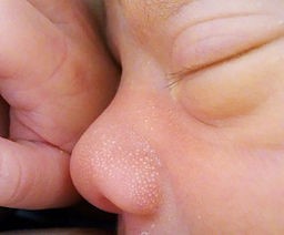
Milia are superficial, uniform, pearly-white to yellowish, domed lesions measuring 1-2 mm in diameter.
Milia most often arise on the face and are particularly prominent on the eyelids and cheeks, but they may occur elsewhere, including the genitalia.
They are usually asymptomatic but may be itchy.
Primary milia
Primary milia in infants occur on the face, especially the cheeks, nose and around the eyes. They may be more widespread on the scalp, face and upper trunk.
Congenital oral inclusion cysts such as those found on the mucosa (Epstein's pearls) and palate (Bohn's nodules) are thought to be the oral counterparts of milia.
Primary milia in children and adults are most often on the eyelids, cheeks, forehead and genitalia. They may clear in a few weeks or persist for months or longer.
Juvenile milia may be present at birth or appear later in life. They may be associated with other genetic disorders.
Milia in a newborn
Milia en plaque3
The small papules arise on a distinct, erythematous plaque in the postauricular area, unilaterally or bilaterally.
They are usually seen behind the ears, but may be on the eyelids, cheeks or on the jaw. Submandibular plaques and lesions on the pinna have been reported.4
They especially affect middle-aged women and may be associated with other skin disease - eg, pseudoxanthoma elasticum, discoid lupus erythematosus, lichen planus.2
Multiple eruptive milia
Crops of milia appear over a few weeks to months.5 Most often, they affect the face, upper arms and upper trunk.2
Secondary milia1
These are found anywhere on the body, depending on the predisposing condition:
Disease: post-bullous, typically epidermolysis bullosa and porphyria cutanea tarda but can also occur after other skin conditions - for example, contact dermatitis or lichen sclerosis.
Medication associated with milia includes topical steroids, 5-fluorouracil, ciclosporin and penicillamine.
Traumatic abrasions are a common cause of milia in children. They can also occur following, for example, second-degree burns, dermabrasion or radiotherapy.
חקירות
The clinical appearance is diagnostic in simple milia and no further investigations are required.
In elderly people with sun-damaged skin, a biopsy may be needed to exclude nodular elastosis of the skin (Favre-Racouchot syndrome). If milia en plaque are suspected, a biopsy is prudent to confirm the diagnosis.
אבחנה מבדלת2
אקנה וולגריס Closed comedones are more cream than white. They also usually have a small punctum as well as being associated with open (black) comedones.
Syringomas are small papules on lower eyelids and are skin-coloured
קסנתלזמה are yellow, flat plaques over the upper or lower eyelids.
Trichoepithelioma.
Follicular (or alopecia) mucinosis.
Milia treatment
Usually no treatment is required.
Some milia may be removed with a needle, as they often shell out easily (this doesn't require anaesthetic).6
Topical peeling agents do not work.
תחזית
They are harmless but can be unsightly. They tend to resolve within a few weeks in infants but may persist in older children and adults.
When to refer milia
For confirmation of diagnosis or reassurance. It is worth referring patients with suspected milia en plaque.
עדכונים בלעדיים לאנשי מקצוע בתחום הבריאות
הישאר מעודכן עם העדכונים הקליניים האחרונים, תובנות מקצועיות והנחיות מבוססות ראיות. הניוזלטר של Patient Pro אוסף תוכן חיוני לאנשי מקצוע בתחום הבריאות - נשלח ישירות לתיבת הדואר הנכנס שלך.
על ידי הרשמה אתה מקבל את שלנו מדיניות הפרטיות שלנו. באפשרותך לבטל את המנוי בכל עת. לעולם לא נמכור את הנתונים שלך.
קריאה נוספת והפניות
- O'Connor NR, McLaughlin MR, Ham P; עור תינוקות: חלק א'. פריחות נפוצות. רופא משפחה אמריקאי. 1 בינואר 2008;77(1):47-52.
- Berk DR, Bayliss SJ; Milia: a review and classification. J Am Acad Dermatol. 2008 Dec;59(6):1050-63. Epub 2008 Sep 25.
- Milium, milia; DermNet NZ
- Martins LE, Werner B, Fonseca GP; Milia en plaque. An Bras Dermatol. 2010 Nov-Dec;85(6):895-8.
- Hallaji Z, Akhyani M, Jamshidi S, et al; Bilateral retro-auricular milia en plaque: a case report and review of the literature. Dermatol Online J. 2010 Jan 15;16(1):12.
- Batra P, Tsou HC, Warycha M, et al; Multiple eruptive milia. Dermatol Online J. 2009 Aug 15;15(8):20.
- Thami GP, Kaur S, Kanwar AJ; Surgical Pearl: Enucleation of milia with a disposable hypodermic needle. J Am Acad Dermatol. 2002 Oct;47(4):602-3.
אודות המחברצפה בפרופיל המלא
עודכן על ידי

Dr Colin Tidy, MRCGP
רופא כללי, מחבר רפואי
MBBS, MRCGP, MRCP (Paediatrics), DCH
ד"ר קולין טיידי הוא רופא ב-NHS, הממוקם באוקספורדשייר.
אודות המבקרצפה בפרופיל המלא

Dr Hayley Willacy, FRCGP
רופא כללי, מחבר רפואי
MBChB (1992), DRCOG, DFFP, MRCOG (Part 1) MRCGP (2007), DFSRH (2013), MSc - medical education (2020)
ד"ר היילי וילאסי הייתה רופאה כללית ב-NHS שעבדה בצפון מערב אנגליה, ופרשה מעבודתה הקלינית בשנת 2022 לאחר 30 שנות עבודה.
גישה פשוטה לטיפולים ושירותי בריאות אמינים באינטרנט.
Common conditions
Get help with common conditions through the NHS, without a GP appointment.





עוד בדרמטולוגיה
- בלניטיס
- נבוס כחול
- כתם קמפבל דה מורגן
- פילריאזיס עורי
- נדידת זחלים בעור
- Erythema induratum
- אריתמה נודוזום
- ליכן פלאנוס
- תסמונת ליפודיסטרופיה
- גושים
- מלנומה ממאירה של העור
- מולוסקום קונטגיוזום
- זיהומי עור מיקובקטריאליים
- אודם בכפות הידיים
- היפרפיגמנטציה לאחר דלקת של העור
- פצעי לחץ
- תסמונת סטיבנס-ג'ונסון
- תסמונת סטרג'-ובר
- כוויית שמש
- נמק אפידרמלי רעיל