לחץ ורידי ג'וגולרי
נבדק על ידי Dr Laurence Knottעודכן לאחרונה על ידי Dr Colin Tidy, MRCGPעודכן לאחרונה 20 דצמבר 2021
עומד בהנחיות העריכה של Patient
- הורדהורד
- שתף
- Language
- דיון
- גרסת שמע
- הוסף למקורות מועדפים בגוגל
אנשי מקצוע רפואיים
מאמרי עיון מקצועיים מיועדים לשימוש של אנשי מקצוע בתחום הבריאות. הם נכתבים על ידי רופאים מבריטניה ומבוססים על ראיות מחקריות, הנחיות בריטיות ואירופאיות. ייתכן שתמצא אחד מהמאמרים שלנו מאמרי הבריאות יותר שימושי.
What is jugular venous pressure?
Jugular venous pressure (JVP) provides an indirect measure of central venous pressure. The internal jugular vein connects to the right atrium without any intervening valves - thus acting as a column for the blood in the right atrium. The JVP consists of certain waveforms and abnormalities of these can help to diagnose certain conditions1 . Unfortunately, detection of these abnormalities and even the JVP itself, can be difficult and has also been superseded by other diagnostic methods.
How to examine jugular venous pressure1
Use the right internal jugular vein (IJV).
The patient should be at a 45° angle.
The patient's head should be turned slightly to the left.
If possible, have a tangential light source that shines obliquely from the left.
Locate the surface markings of the IJV - this runs from the medial end of the clavicle to the ear lobe, under the medial aspect of the sternocleidomastoid.
Locate the JVP - look for the double waveform pulsation (palpating the contralateral carotid pulse will help).
Measure the level of the JVP by measuring the vertical distance between the sternal angle and the top of the JVP. Measure the height - usually less than 3 cm.
Waveforms of jugular venous pressure
Jugular venous pulse
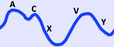
© By Ecgtocardiology, via Wikimedia Commons
מידע חשוב |
|---|
Waves1 כ - presystolic; produced by right atrial contraction. c - bulging of the tricuspid valve into the right atrium during ventricular systole (isovolumic phase). v - occurs in late systole; increased blood in the right atrium from venous return. Descents x - a combination of atrial relaxation, downward movement of the tricuspid valve and ventricular systole. y - the tricuspid valve opens and blood flows into the right ventricle. |
The a and v waves can be identified by timing the double waveform with the opposite carotid pulse. The a wave will occur just before the pulse and the v wave occurs towards the end of the pulse. Distinguishing the c wave, x and y descents is an almost impossible task.
How to differentiate a jugular venous pulse from the carotid pulse
The jugular venous pulse is:
Not palpable.
Obliterated by pressure.
Characterised by a double waveform.
Variable with respiration - it decreases with inspiration.
Enhanced by the hepatojugular reflux (see below).
Hepatojugular reflux (abdominojugular reflux sign)2
This can help to confirm that the pulsation is caused by the JVP.
In the classic test for hepatojugular reflux, firm pressure is applied to the right upper quadrant using the palm of the hand. It has been realised that pressure anywhere over the abdomen will produce the same result (abdominojugular reflux sign). Pressure over the peri-umbilical region is the usual method and may be more appropriate in patients with a tender liver.
A transient increase in the JVP will be seen in normal patients.
There may be a delayed recovery back to baseline which is more marked in right ventricular failure.
Raised jugular venous pressure causes
Heart failure.
Constrictive pericarditis (JVP increases on inspiration - called Kussmaul's sign).
Cardiac tamponade.
Fluid overload - eg, renal disease.
Superior vena cava obstruction (no pulsation).
Jugular venous pressure abnormalities1 3
Abnormalities of the a wave
It disappears in atrial fibrillation.
Large a waves occur in any cause of right ventricular hypertrophy (pulmonary hypertension and pulmonary stenosis) and tricuspid stenosis.
Extra large a waves (called cannon waves) in complete heart block and ventricular tachycardia.
Prominent v waves
Tricuspid regurgitation - called cv or v waves and occurring at the same time as systole (a combination of v wave and loss of x descent); there may be earlobe movement.
Slow y descent
Tricuspid stenosis.
Right atrial myxoma.
Steep y descent
Right ventricular failure.
Constrictive pericarditis.
Tricuspid regurgitation.
(The last two conditions have a rapid rise and fall of the JVP - called Friedreich's sign.)
Prognostic use of jugular venous pressure
Elevated JVP in patients with heart failure is associated with an increased risk of hospital admission, death and subsequent hospitalisation for heart failure4 . Therefore, appreciation of this sign can be clinically helpful.
עדכונים בלעדיים לאנשי מקצוע בתחום הבריאות
הישאר מעודכן עם העדכונים הקליניים האחרונים, תובנות מקצועיות והנחיות מבוססות ראיות. הניוזלטר של Patient Pro אוסף תוכן חיוני לאנשי מקצוע בתחום הבריאות - נשלח ישירות לתיבת הדואר הנכנס שלך.
על ידי הרשמה אתה מקבל את שלנו מדיניות הפרטיות שלנו. באפשרותך לבטל את המנוי בכל עת. לעולם לא נמכור את הנתונים שלך.
קריאה נוספת והפניות
- Chua Chiaco JM, Parikh NI, Fergusson DJ; The jugular venous pressure revisited. Cleve Clin J Med. 2013 Oct;80(10):638-44. doi: 10.3949/ccjm.80a.13039.
- Brostoff JM; Re-examining examination: misconceptions in clinical medicine. J R Soc Med. 2009 Jan;102(1):11-5. doi: 10.1258/jrsm.2008.080330.
- Witteles R; Neck Veins & Wave Forms, Stanford Medicine 25
- Drazner MH, Rame JE, Stevenson LW, et al; Prognostic importance of elevated jugular venous pressure and a third heart sound in patients with heart failure. N Engl J Med. 2001 Aug 23;345(8):574-81.
אודות המחברצפה בפרופיל המלא

Dr Colin Tidy, MRCGP
רופא כללי, מחבר רפואי
MBBS, MRCGP, MRCP (Paediatrics), DCH
ד"ר קולין טיידי הוא רופא ב-NHS, הממוקם באוקספורדשייר.
אודות המבקרצפה בפרופיל המלא

Dr Laurence Knott
רופא כללי, מחבר רפואי
תואר ראשון (בהצטיינות) בביוכימיה, דוקטור לרפואה
ד"ר לורנס קנוט הוסמך בשנת 1973 ויש לו ניסיון נרחב כרופא כללי.
היסטוריית המאמר
המידע בעמוד זה נכתב ונבדק על ידי קלינאים מוסמכים.
המאמר זמין גם ב אנגלית, גרמנית, ספרדית, צרפתית, איטלקית, פורטוגזית, הינדי, עברית, ערבית, and שוודית.
הסקירה הבאה מתוכננת ל: 19 דצמבר 2026
20 דצמבר 2021 | הגרסה האחרונה

שאלו, שתפו, התחברו.
עיין בדיונים, שאל שאלות ושתף חוויות במאות נושאים בריאותיים.

מרגיש לא טוב?
הערך את הסימפטומים שלך באינטרנט בחינם
עוד בהיסטוריה ובבדיקה
- מסות בטניות
- כאבי גב אצל ילדים
- ורטיגו תנוחתי פרוקסיזמלי שפיר
- אישור פטירה
- התקף ראשון
- היסטוריה ובדיקה של מערכת העיכול
- היסטוריה ובדיקה של מערכת השתן והמין - נשים
- היפרוונטילציה
- הרניות מפשעתיות
- התעלפות בעת מתן שתן
- התכווצויות שרירים
- פרוקטלגיה פוגקס וכאב אנאלי
- מחלת ריינו
- תסמונת סינוס חולה
- טכיקרדיה על-חדרית במבוגרים
- כוריאה של סידנהם
- Tenesmus
- התרחבות אצבעות הידיים והרגליים
- רעידה
- דליפת שתן