בדיקה נוירולוגית של הגפיים התחתונות
נבדק על ידי Dr Philippa Vincent, MRCGPעודכן לאחרונה על ידי Dr Toni Hazell, FRCGPעודכן לאחרונה 20 במאי 2026
עומד בהנחיות העריכה של Patient
- הורדהורד
- שתף
- Language
- דיון
- Ask Patient
- גרסת שמע
- הוסף למקורות מועדפים בגוגל
אנשי מקצוע רפואיים
מאמרי עיון מקצועיים מיועדים לשימוש של אנשי מקצוע בתחום הבריאות. הם נכתבים על ידי רופאים מבריטניה ומבוססים על ראיות מחקריות, הנחיות בריטיות ואירופאיות. ייתכן שתמצא אחד מהמאמרים שלנו מאמרי הבריאות יותר שימושי.
יש יותר מדרך אחת לבצע בדיקה נוירולוגית, ורופא צריך לפתח את הטכניקה האישית שלו. טכניקה לקויה לא תצליח להפיק סימנים או תייצר תוצאות שגויות.
ראה את הנפרד היסטוריה ובדיקה נוירולוגית מאמר המכסה את העקרונות הבסיסיים של בדיקה וטכניקה.
Inspection of the lower limbs1
Note whether there is any damage to the feet. Neuropathic ulcers and neuropathic joints develop because of a lack of pain perception. Pain is a protective mechanism. When inspecting the motor system, the following points should be assessed:
Note the resting posture. Establish whether there is unusual rotation or posture of a joint. Note whether anything about the patient is asymmetrical.
Look for muscle wasting or hypertrophy: note whether it is focal or diffuse.
חפש תנועות לא רצוניות כמו רעד, טיקים, קפיצות מיוקלוניות, כוראה או אתטוזיס.
Look for muscle fasciculation (a sign of lower motor neurone disease process). These are subcutaneous twitches over a muscle belly at rest. Tapping the belly may stimulate fasciculation.
Lower limbs examination of the sensory system
Examination of the lower limbs may be performed more easily with the patient lying on a couch.
Examination of each of the sensory modalities1
מגע קל
השתמשו במגע קל של אצבע, חתיכת צמר גפן או חתיכת נייר טישו.
חשוב לגעת ולא ללטף, שכן תחושה נעה, כמו שפשוף וגרד, מועברת לאורך מסלולי הכאב.
בקש מהמטופל לעצום את עיניו ולהגיד לך מתי הוא מרגיש שאתה נוגע בו.
השווה כל גפה באותה תנוחה.
שמור על תזמון לא סדיר של כל מגע כדי למנוע ציפייה מצד המטופל.
A logical progression is required. You may want to start testing over the groin and to move down the front of the leg and up the posterior side as this moves progressively from L1 to S3 dermatomes.
ציין כל אזור של היפואסתזיה או דיסאסתזיה.
מגע חד (דקירה)
בדוק באמצעות סיכה חד פעמית ייעודית. מחט היפודרמית חד פעמית חדה מדי.
השתמש באזור הסטרנום כדי לקבוע קו בסיס לחדות לפני שתתחיל.
Follow the same progression as with light touch with the patient's eyes closed, comparing both lower limbs.
בקש מהמטופל לדווח על היפואסתזיה (מרגיש קהה יותר) או היפראסתזיה (מרגיש חד יותר).
טמפרטורה
לעיתים קרובות מתעלמים מזה, אבל זה יכול להיות חשוב.
An easy and practical approach is to touch the patient with a tuning fork, as the metal feels cold.
השווה את איכות תחושת הטמפרטורה על הזרועות, הפנים, הגו, הידיים, הרגליים וכפות הרגליים.
ניתן להשתמש במיכלים של מים חמים וקרים להערכה מדויקת יותר. בקש מהמטופל להבחין בין חם לקר באזורים שונים של העור בעיניים עצומות.
תחושת מיקום המפרק (פרופריוספציה)
Test at the interphalangeal joint of the big toe.
Hold the proximal phalanx with one thumb and finger and hold the medial and lateral sides of the distal phalanx with the other. Move the distal phalanx up and down, showing the patient the movement first.
בקש מהמטופל לעצום את עיניו ולהזיז את החלק הדיסטלי של האצבע למעלה ולמטה באופן אקראי. בקש מהמטופל לומר לך את כיוון התנועה בכל פעם.
Test on both feet.
If there is an abnormality, move backwards to the metatarsophalangeal joint and so on until joint position sense is normal.
תחושת רטט
השתמש במזלג כיוון של 128 הרץ וודא שהמזלג רוטט.
הנח אותו על עצם החזה כדי להתחיל כך שהמטופל יוכל להרגיש את התחושה.
Then place it on the big toe.
If no vibration is sensed, move backwards to the bony malleolus of the ankle, the tibial shaft and tuberosity and the anterior iliac crest.
לבקש מהמטופל לומר לך מתי המזלג המכוון מפסיק לרטוט יכול להיות מועיל אם יש ספק שהתחושה שלהם לרטט תקינה.
2-point discrimination
This test is not usually performed on the soles of the feet because the distinguishing distance is usually much greater than that on the fingers.
פירוש הממצאים
The site of any lesion can be determined by looking at the pattern of any dysfunction found. The dermatomal (segmental) and peripheral nerve innervation is labelled below.
בדיקה נוירולוגית
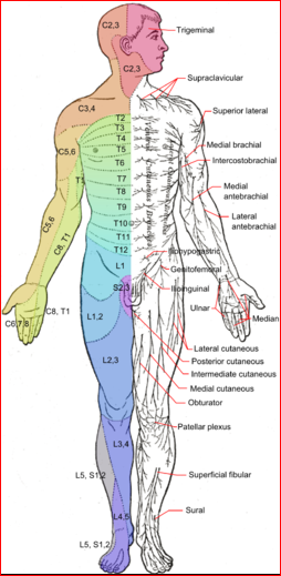
All of the sensory modalities can be affected in peripheral neuropathies and nerve injuries, radiculopathy due to disc lesions and spinal injuries.
אם עצב יחיד או שורש תחושתי מושפע, כל המודלים התחושתיים יכולים להיות מופחתים.
If there is a spinal cord lesion, there may not be equal diminution across all of the sensory modalities: light touch, vibration and joint position sense may remain intact while sharp touch and temperature are lost. This is because the lateral spinothalamic pathways may be damaged while the dorsal columns remain intact.
Problems with joint position sense or vibration usually occur distally first. Joint position sense is also required for balance, gait and co-ordination.
תחושת הרטט יכולה להיעלם לפני תחושת מיקום המפרק בנוירופתיה פריפרית או מיאלופתיה המשפיעה על העמודות האחוריות.
Vibration sense over the ankles is often diminished or lost in elderly people with no apparent neurological lesion.
החלקים הדיסטליים של הגפיים נוטים להיות מושפעים בפולינוירופתיה, כאשר הרגליים בדרך כלל מעורבות לפני הידיים. נוצר אפקט של 'כפפה וגרב'.
Lower limbs examination of the motor system2
גוון
Tone is the resistance felt when a joint is moved passively through its normal range of movement. Hypertonia is found in upper motor neurone lesions; hypotonia is found in lower motor neurone lesions and cerebellar disorders.
Clonus is rhythmic and involuntary muscle contraction that can be provoked by stretching a group of muscles.
Test tone:
Ask the patient to let their legs 'go floppy'.
Internally and externally rotate the 'floppy' leg. Assess for any increased or reduced tone.
Then lift the knee off the bed with one of your hands. Note whether the ankle raises off the bed as well, signifying increased tone.
Test for ankle clonus:
Flex the patient's knee, resting the ankle on the bed.
Dorsiflex the foot quickly and keep the pressure applied.
You will be able to see the foot moving up and down if clonus is present.
כוח3
נדרשת הערכה מקיפה של הכוח.
The Medical Research Council (MRC) has a recommended grading system for power (see table). It has been shown to have good reliability although doubts have been expressed over the wide range of grade.4
בדיקת כוח השרירים הידנית מדורגת באופן שונה עם 4 - טוב: טווח תנועה מלא נגד כוח הכבידה עם התנגדות מתונה, ו-5 - נורמלי: טווח תנועה מלא נגד כוח הכבידה עם התנגדות מרבית.
Get the patient to contract the muscle group being tested and then you as the examiner try to overpower that group.
בדוק את הדברים הבאים:
Hip flexion, extension, adduction and abduction.
Knee flexion and extension.
Foot dorsiflexion, plantar flexion, eversion and inversion.
Toe plantar flexion and dorsiflexion.
סולם MRC לכוח שרירים
0 | לא נראית התכווצות שרירים. |
1 | התכווצות השריר נראית לעין אך אין תנועה של המפרק. |
2 | תנועה פעילה של המפרק אפשרית כאשר הכבידה מבוטלת. |
3 | תנועה יכולה להתגבר על כוח הכבידה אך לא על התנגדות הבוחן. |
4 | קבוצת השרירים יכולה להתגבר על כוח הכבידה ולנוע כנגד התנגדות מסוימת מצד הבוחן. |
5 | כוח מלא ונורמלי כנגד התנגדות. |
רפלקסים של גידים עמוקים
ודא שהמטופל נינוח ורגוע ושאתה יכול לראות את השריר הנבדק.
השתמש בפטיש גידים כדי להכות בגיד של השריר ולחפש התכווצות שרירים.
השווה בין שני הצדדים.
רפלקסים יכולים להיות היפראקטיביים (+++), נורמליים (++), איטיים (+) או חסרים (-). ± משמש כאשר הרפלקס קיים רק בחיזוק (ראה למטה).
In the lower limbs:
Test the knee jerk (L3, L4): flex the patient's knee and support it by using one of your hands in their popliteal fossa. Elicit the reflex by tapping just below the patella.
Test the ankle jerk (S1): with the patient lying down, flex their knee and dorsiflex their ankle, at the same time rotating their leg slightly laterally. Elicit the reflex by tapping over the Achilles tendon just above the heel. If this is difficult to elicit, an alternative method is to ask the patient to kneel on a chair, facing the back of the chair, so that their feet are dangling off the seat of the chair. Tap over the same area in this position.
If a reflex is difficult to elicit, try 'reinforcement' (the Jendrassik manoeuvre). Ask the patient to flex their fingers and interlock them with one palm facing upwards and the other facing downwards. Then ask them to try to pull their fingers apart just before you strike the tendon.
פרשנות
נגעים במערכת העצבים העליונה בדרך כלל גורמים להיפררפלקסיה.
נגעים בנוירונים מוטוריים תחתונים בדרך כלל גורמים לתגובה מופחתת או נעדרת.
It may be normal to have reduced or absent ankle reflexes in some elderly people, although the frequency and significance of this is disputed.
Isolated loss of a reflex can point to a radiculopathy affecting that segment - eg, loss of ankle jerk if there is an S1 disc prolapse.
Superficial tendon reflexes
In the lower limbs, this is the plantar response.
To elicit this, the patient should be lying down with their legs extended.
Use a blunted point and run this along the lateral border of the foot, starting at the heel and moving towards the big toe.
Stop on the first movement of the big toe.
An extensor plantar response (upgoing big toe) is pathological and signifies an upper motor neurone lesion.
בדיקת תיאום
The cerebellum helps in the co-ordination of voluntary, automatic and reflex movement. Tests of cerebellar function in the lower limbs require normal power, tone and sensation to be valid and include:
The heel-shin test:
Ask the patient to lift one of their legs and flex it at the knee, keeping the other leg straight.
They should then place the heel of the flexed leg on the knee of the other leg and run it down the shin towards the ankle and back again towards the knee.
Ask them to repeat this a number of times.
The heel-toe test:
This tests balance mechanisms that rely on the cerebellar, vestibular and proprioceptive systems.
The patient either needs to be barefoot or wearing flat shoes.
They should walk in a straight line so that the heel of the second foot touches the toes of the first foot. This should be repeated so that the heel of the first foot then touches the toes of the second foot, etc, each time with the patient moving forward.
Look at how well they are able to perform this. Is there any staggering which would suggest a lesion of the cerebellum?
Romberg's test:
This also tests balance mechanisms that rely on the cerebellar, vestibular and proprioceptive systems.
Ask the patient to keep their eyes open and stand with their feet together, arms by their sides.
Then ask them to maintain this position when they close their eyes.
Patients who have cerebellar lesions often cannot stand in this position, even with their eyes open. If balance is only lost when the eyes are closed, this signifies a proprioceptive or vestibular lesion.
Be ready to catch the patient by standing behind.
Examining the lower limbs and gait
No neurological examination, especially of the lower limbs, is complete without observing gait.
This is an enormous topic and there are separate articles discussing הליכה לא תקינה ו הפרעות הליכה בילדים.
Also, watch the patient as they rise from the chair to walk and note any abnormality of movement.
עדכונים בלעדיים לאנשי מקצוע בתחום הבריאות
הישאר מעודכן עם העדכונים הקליניים האחרונים, תובנות מקצועיות והנחיות מבוססות ראיות. הניוזלטר של Patient Pro אוסף תוכן חיוני לאנשי מקצוע בתחום הבריאות - נשלח ישירות לתיבת הדואר הנכנס שלך.
על ידי הרשמה אתה מקבל את שלנו מדיניות הפרטיות שלנו. באפשרותך לבטל את המנוי בכל עת. לעולם לא נמכור את הנתונים שלך.
קריאה נוספת והפניות
- החברה לנוירולוגיה בטיפול ראשוני
- בדיקה נוירולוגית; Geeky medics (accessed November, 2021)
- בדיקה נוירולוגית; בדיקה רפואית אוקספורד (OME)
- Shahrokhi M, Asuncion RMD; בדיקה נוירולוגית
- Naqvi U, Sherman Al; דירוג כוח שרירים
אודות המחברצפה בפרופיל המלא
עודכן על ידי

Dr Toni Hazell, FRCGP
MBBS, BSc, FRCGP, DFSRH, Dip GU med, DRCOG, DCH (London, UK, 2000)
ד"ר טוני הייזל סיימה את לימודיה בבית הספר לרפואה של בית החולים סנט מרי ועשתה את התמחותה בבית החולים נורת'וויק פארק.
אודות המבקרצפה בפרופיל המלא

Dr Philippa Vincent, MRCGP
רופא כללי, מחבר רפואי
MB BS, Bsc, MRCGP (2000), DCH, DFSRH, DRCOG
ד"ר Philippa וינסנט הוא רופא משפחה ב-NHS העובד בצפון לונדון.
גישה פשוטה לטיפולים ושירותי בריאות אמינים באינטרנט.
Common conditions
Get help with common conditions through the NHS, without a GP appointment.





עוד בהיסטוריה ובבדיקה
- בדיקות טרום לידתיות ואבחון הריון
- ניקור מיימת
- שיתוק בולברי ושיתוק פסאודובולברי
- כאב חזה מסוג לב המופיע בטיפול ראשוני
- דלקת אוזן תיכונה כרונית מוגלתית
- תרדמת
- עצירות במבוגרים
- התייבשות אצל ילדים
- בקע פמורלי
- לימפדנופתיה כללית
- דפיקות לב
- צהבת
- אובדן חשק מיני
- בדיקת שישה שבועות לאחר לידה לאם
- השמנת יתר בילדים
- כף רגל כואבת
- פרסביאקוזיס
- מניעת נפילות בקרב קשישים
- כאב ברביע הימני העליון
- נבוס עכביש