סריקת רדיונוקלידים
Isotope scan
נבדק על ידי Dr Krishna Vakharia, MRCGPעודכן לאחרונה על ידי ד"ר רייצ'ל הדסון, MRCGPעודכן לאחרונה 24 Aug 2023
עומד בהנחיות העריכה של Patient
- הורדהורד
- שתף
- Language
- דיון
- Ask Patient
- גרסת שמע
- הוסף למקורות מועדפים בגוגל
A radionuclide scan is a way of imaging bones, organs and other parts of the body by using a small dose of a radioactive chemical. There are different types of radionuclide chemical. The one used depends on which organ or part of the body is to be scanned.
הערה: המידע להלן הוא מדריך כללי בלבד. הסידורים והאופן שבו מבוצעים הבדיקות עשויים להשתנות בין בתי חולים שונים. תמיד יש לפעול לפי ההנחיות שניתנו על ידי הרופא שלך או בית החולים המקומי.
במבט חטוף
סריקת רדיונוקלידים משתמשת בכמות קטנה של חומר רדיואקטיבי כדי ליצור תמונות של פנים גופך.
זה יכול לעזור בזיהוי מצבים כמו סרטן, זיהום, קרישי דם, או בהערכת תפקוד איברים.
הכימיקל בדרך כלל נכנס לגופך באמצעות הזרקה או בליעה, ומצלמה מיוחדת מזהה את האותות שלו.
ההכנה עשויה לכלול שתיית נוזלים או הפסקת תרופות מסוימות.
כמות הקרינה המשמשת היא קטנה ונחשבת בדרך כלל לבטוחה.
ספרי לרופא שלך אם את בהריון או עשויה להיות, שכן נשים בהריון לא צריכות לעבור את הסריקות הללו.
Why do you have a radionuclide scan?
A radionuclide scan may be done for all sorts of reasons. For example:
A bone scan is a common type. A radionuclide is used which collects in areas where there is a lot of bone activity (where bone cells are breaking down or repairing parts of the bone). So a bone scan is used to detect areas of bone where there is cancer, infection, or damage. These areas of activity are seen as 'hot spots' on the scan picture. See the separate leaflet called Bone Scan for more details.
A kidney scan can assess how well a kidney is working (as the radionuclide chosen is taken up by kidney cells and passes into the urine). So, the scan can detect scars on the kidney and how well urine drains from the kidney to the bladder. See the separate leaflet called DMSA Scan for more details.
Lung perfusion scan (also called a 'VQ scan') can detect blood clots in the lungs (pulmonary embolism).
A heart scan can assess blood flow to the heart muscle. Areas of poor blood flow to the heart muscle do not 'take up' the radionuclide very well and this will be shown in the picture.
A thyroid scan may be done to assess cases of overactive thyroid (hyperthyroidism). For example, some nodules (small 'lumps') are sometimes a focus of overactivity and will show as 'hot spots' on the picture. See the separate leaflet called Thyroid Scans and Uptake Tests for more details.
Lacrimal scintigraphy is done to test the function of tear ducts (lacrimal ducts.) The radionuclide is given as eye drops.
Lymphoscintigraphy is done to check the drainage of the בלוטות לימפה in people with a type of swelling of the legs, called lymphoedema.
There are various other types of radionuclide tests.
What preparation do I need?
The preparation needed is usually very little. It will depend on which type of scan you are having. Your local hospital should give you specific information to help you prepare for these tests.
For some types of scan, you may be asked to have lots to drink to help to flush the radionuclide through your body.
For some types of scan you may also be asked to empty your bladder of urine before the scanning begins.
For some scans, such as thyroid scans, you may be instructed to stop certain medications for some time before the scan.
As these tests involve a small amount of radiation, pregnant women should not have them.
הערה: let your doctor know if you are, or think you could be, pregnant. You should also let your doctor know if you are breastfeeding.
What happens during a radionuclide scan?
The procedures for the different types of radionuclide scans are different. Information about your scan should be sent to you with the appointment.
Depending on the type of scan you have, you usually either swallow a small quantity of radionuclide, or it is injected into a vein in your arm.
It then takes some time - sometimes several hours (depending on what is being scanned) - for the radionuclide to travel to the target organ or tissue, and to be 'taken' into the active cells.
So, after receiving the radionuclide you may have a wait of a few hours. You may be able to go out and come back to the scanning room later in the day.
When it is time to do the scanning, you usually lie on a couch while the gamma camera detects the gamma rays coming from your body.
Gamma camera
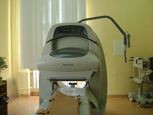
The computer turns the information into a picture. You need to lie as still as possible whilst each picture is taken (so it is not blurred). Some pictures can take 20 minutes or more to expose.
The number of pictures taken and the time interval between each picture vary depending on what is being scanned. Sometimes only one picture is needed. However, for some scans (such as bone scans or heart scans), two or more pictures are needed. Each picture may be taken several hours apart. So, the whole process can take several hours.
What happens after a radionuclide scan?
Radionuclide scans do not generally cause any side-effects.
Uncommon side-effects from radionuclides may include flushing, racing heart and בחילה but these are short-lived because they are flushed out of your system quickly.
Through the natural process of radioactive decay, the small amount of radioactive chemical in your body will lose its radioactivity over time. It may also pass out of your body through your urine or poo during the first few hours or days following the test.
You may be instructed to take special precautions after urinating, to flush the toilet twice and to wash your hands thoroughly. You may be advised to drink plenty of water to help flush the chemicals out of your system.
If you have contact with children or pregnant women you should let your doctor know. Although the levels of radiation used in the scan are small, they may advise special precautions. Your hospital should give you more advice on this.
Are there risks with radionuclide scans?
The term 'radioactivity' may sound alarming. But, the radioactive chemicals used in radionuclide scans are considered to be safe, and they leave the body quickly in the urine. The dose of radiation that your body receives is very small.
In many cases, the level of radiation involved is not much different to a series of a few normal X-rays. However:
As with any other types of radiation (such as X-ray), there is a small risk that the gamma rays may affect an unborn child. So, tell your doctor if you are pregnant or if you may be pregnant.
Rarely, some people have an allergic reaction to the injected chemical. Tell your doctor if you are allergic to iodine.
Theoretically, it is possible to receive an overdose when the chemical is injected. This is very rare.
How does a radionuclide scan work?
A radionuclide (sometimes called a radioisotope or isotope) is a chemical which emits a type of radioactivity called gamma rays. A tiny amount of radionuclide is put into the body, usually by an injection into a vein. Sometimes it is breathed in, or swallowed, or given as eye drops, depending on the test.
There are different types of radionuclides. Different ones tend to collect or concentrate in different organs or tissues. So, the radionuclide used depends on which part of the body is to be scanned.
For example, if radioactive iodine is injected into a vein it is quickly taken up into the tissues of the thyroid gland. So, it is used to scan the thyroid gland.
Cells which are most 'active' in the target tissue or organ will take up more of the radionuclide. So, active parts of the tissue will emit more gamma rays than less active or inactive parts.
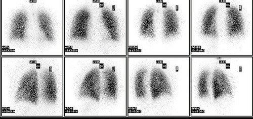
Gamma rays are similar to X-rays and are detected by a device called a gamma camera. The gamma rays which are emitted from inside the body are detected by the gamma camera, are converted into an electrical signal and sent to a computer.
The computer builds a picture by converting the differing intensities of radioactivity emitted into different colours or shades of grey. This is seen below in a lung perfusion scan.
Lung perfusion scan
Alternatively areas of the target organ or tissue which emit lots of gamma rays may be shown as red spots ('hot spots') on the picture on the computer monitor.
Areas which emit low levels of gamma rays may be shown as blue ('cold spots'). Various other colours may be used for 'in between' levels of gamma rays emitted.
בחירות המטופלים עבור הדמיה
בדיקות וחקירות
בדיקת DMSA
סריקת DMSA משתמשת בחומר רדיואקטיבי כדי ליצור תמונות מיוחדות של הכליות. היא יכולה לעזור להראות האם הכליות פגועות או מצולקות. הערה: המידע למטה הוא מדריך כללי בלבד. הסידורים והאופן שבו מבוצעים הבדיקות עשויים להשתנות בין בתי חולים שונים. תמיד יש לעקוב אחר ההוראות שניתנו על ידי הרופא שלך או בית החולים המקומי.
מאת ד"ר דאג מקקנזי, MRCGP

בדיקות וחקירות
בדיקות בריום
בדיקות בריום משמשות כדי לעזור לראות את קווי המתאר של חלקים שונים במערכת העיכול. אלה כוללים את הוושט, הקיבה, המעי הדק והמעי הגס. בדיקות רנטגן בריום נעשות פחות נפוצות בימינו. כיום אנו בדרך כלל בודקים את המעי עם טלסקופ גמיש (אנדוסקופיה או קולונוסקופיה). עם זאת, עדיין יש מקום לבדיקות בריום כדי לעזור להעריך בעיות שונות במערכת העיכול. הערה: המידע להלן הוא מדריך כללי בלבד. הסידורים והאופן שבו מבוצעות הבדיקות עשויים להשתנות בין בתי חולים שונים. תמיד יש לעקוב אחר ההוראות שניתנו על ידי הרופא שלך או בית החולים המקומי.
מאת ד"ר רוזלין אדלמן, MRCGP
שאלות נפוצות
מהו רדיונוקליד?
רדיו-נוקליד, הידוע גם כרדיואיזוטופ או איזוטופ, הוא חומר כימי הפולט קרני גמא. כמות קטנה של חומר זה מוחדרת לגוף כדי לאפשר למצלמת גמא ליצור תמונות של איברים פנימיים או רקמות. רדיו-נוקלידים שונים משמשים בהתאם לאיבר או לרקמה הספציפיים הנבדקים.
האם יש רדיונוקלידים ספציפיים לחלקי גוף שונים?
כן, ישנם סוגים שונים של רדיונוקלידים, וכל אחד מהם נוטה להצטבר באיברים או ברקמות ספציפיות. הבחירה ברדיונוקליד תלויה באיזה חלק של הגוף יש לסרוק. לדוגמה, יוד רדיואקטיבי משמש לסריקות של בלוטת התריס מכיוון שהוא נספג בקלות ברקמות התריס.
כיצד מפענחים את התמונות מסריקת רדיונוקלידים?
המצלמה הגמאית מזהה את קרני הגמא הנפלטות מתוך גופך וממירה אותן לאותות חשמליים עבור מחשב. המחשב יוצר תמונה, תוך שימוש בצבעים שונים או בגוונים של אפור כדי לייצג עוצמות שונות של רדיואקטיביות. "נקודות חמות" (לעיתים מוצגות באדום) מצביעות על אזורים עם פליטת קרני גמא גבוהה, כלומר פעילות גבוהה, בעוד "נקודות קרות" (לעיתים בכחול) מצביעות על פליטה או פעילות נמוכה. צבעים אחרים עשויים לשמש לרמות ביניים.
האם אני יכולה לעבור סריקת רדיונוקלידים אם אני מניקה?
אם את מניקה, עלייך ליידע את הרופא שלך לפני ביצוע סריקת רדיונוקלידים. הם יוכלו לייעץ לך על אמצעי זהירות מיוחדים שעשויים להיות נחוצים.
מה קורה אם יש לי תגובה אלרגית לחומר הכימי שהוזרק במהלך הסריקה?
תגובות אלרגיות לחומר המוזרק הן נדירות. עם זאת, אם יש לך אלרגיות ידועות, במיוחד ליוד, עליך ליידע את הרופא שלך מראש.
האם אחוש באי נוחות כלשהי במהלך תהליך הסריקה עצמו?
במהלך הסריקה עצמה, בדרך כלל תשכב ללא תנועה על ספה בזמן שהמצלמה הגמא מצלמת תמונות. תצטרך להישאר כמה שיותר ללא תנועה כדי למנוע טשטוש של התמונות. חלק מהתמונות יכולות לקחת 20 דקות או יותר לחשיפה.
קריאה נוספת והפניות
- Myocardial perfusion scintigraphy for the diagnosis and management of angina and myocardial infarction; NICE Technology Appraisal Guidance, November 2003 (last updated July 2011)
- Nuclear Medicine; Radiology.info.org (American College of Radiology and Radiological Society of North America)
- Guidelines; British Nuclear Medicine Society
- Gruning T, Drake BE, Freeman SJ; Single-photon emission CT using (99m)Tc-dimercaptosuccinic acid (DMSA) for characterization of suspected renal masses. Br J Radiol. 2014 Jul;87(1039):20130547. doi: 10.1259/bjr.20130547. Epub 2014 May 16.
- Radionuclide bone scans/bone scintigraphy; מחקר סרטן בריטניה
- Myocardial perfusion scans; הקרן הבריטית ללב
אודות המחברצפה בפרופיל המלא
עודכן על ידי

ד"ר רייצ'ל הדסון, MRCGP
רופא כללי ומחבר רפואי
MBChB, MRCGP (2008), BSc (Medical Science), DFSRH, DRCOG, DCH
ד"ר רייצ'ל הדסון, היא רופאה כללית ב-NHS העובדת בצפון מערב אנגליה.
אודות המבקרצפה בפרופיל המלא

Dr Krishna Vakharia, MRCGP
קצין רפואה ראשי לבריאות, Optum UK
MBChB, MRCGP(2013), BMedSci (hons), DFSRH, DRCOG, PGDipDerm (Distn)
ד"ר קרישנה וקאריה היא רופאת משפחה ב-NHS. היא גם בוחנת קבועה לדיפלומה לתואר שני בדרמטולוגיה מעשית באוניברסיטת קרדיף וכן משמשת כקצינת הרפואה הראשית לבריאות ב-Optum UK.
שתפו את מחשבותיכם או חוויותיכם עם אחרים ב קהילות המטופלים שלנו.
גישה פשוטה לטיפולים ושירותי בריאות אמינים באינטרנט.
Common conditions
Get help with common conditions through the NHS, without a GP appointment.





הירשמו לניוזלטר של פיישנט
המנה השבועית שלך של עצות בריאות ברורות ואמינות - נכתבה כדי לעזור לך להרגיש מעודכן, בטוח ובשליטה.
על ידי הרשמה אתה מקבל את שלנו מדיניות הפרטיות שלנו. באפשרותך לבטל את המנוי בכל עת. לעולם לא נמכור את הנתונים שלך.